
Textured Bioplastic Proves Superior to Smooth Bioplastic for Implants
14 October 2026 г. FRC KSC SB RAS
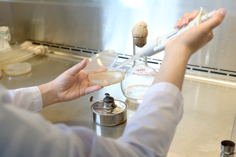
Scientists have found that smooth medical bioplastics are inferior to textured ones. To ensure the cell attachment and growth on an implant, a surface should be textured and have roughness and indentations. The results of the study have been published in the Langmuir Journal.
Polyhydroxyalkanoates (PHAs) are biodegradable polymers synthesized by bacteria. They are biocompatible with living cells and tissues and degrade into non-toxic substances within the body, which makes them ideal candidates for biomedical applications, including implants, sutures, and tissue engineering scaffolds. However, simply producing a biocompatible material or medical device is not enough; cells should be able to attach to and grow on it. This depends on the surface topography on the micro- and nanoscale. Therefore, the texture of the surface device is crucial for use in biomedicine.
Researchers from the ITMO University (St. Petersburg), the Krasnoyarsk Scientific Center of the Siberian Branch of the Russian Academy of Sciences, and the Siberian Federal University in cooperation with colleagues from the Netherlands and the US have determined the optimum thickness and surface topography of PHA polymer films for the cell attachment and growth. The films have been fabricated from a biodegradable PHA polymer selected due to the ease of processing, high biocompatibility, and cost efficiency in large-scale biosynthesis. The films have been obtained using four polymer solutions of varying concentrations; the higher the solution concentration, the thicker and rougher the film. The scientists then seeded C2C12 myoblast cells onto the films. These cells are precursors to muscle tissue and one of the most convenient and widely used models for testing biomaterials.
During the project, the researchers faced the challenge of evaluating suitability of the surface for cells. This is conventionally performed manually: counting cells, taking measurements, and separately describing the surface topography. The process is time-consuming, subjective, and difficult to consistently replicate across experiments. The authors have developed a reproducible algorithm that automates these tasks, segmenting cell images and generating metrics for both the surface topography and cells themselves. In addition, the algorithm links every process stage, from film fabrication and surface characterization to obtaining data on cell behavior, into a single workflow. This approach enables the targeted selection of parameters rather than relying on trial and error. In the development of new polymer implants, the technique is applicable to any human cells.
"Our team from the Infochemistry Scientific Center of the ITMO University has developed an approach for automated evaluation of how cells interact with the polymer films surface. Fluorescence microscopy images of cell are segmented using a convolutional neural network, which is followed by checking the segmentation quality. After that, we extract the key characteristics of cell, including shape, size, and other morphometric features, and compare them with the surface properties established from atomic force and scanning electron microscopy images. This allows us to identify the surface characteristics most strongly linked to the biocompatibility of samples," noted Pavel Zun, Associate Prof. at the Infochemistry Scientific Center, Head of the biomedical computing team at the ITMO University.
Using the proposed algorithm, the researchers monitored how cells behave on the films, specifically, how they attach and proliferate, and how efficiently the film supports tissue growth. It has been found that cells attach better to thicker, rougher films. Surface irregularities, which provide anchor points for cells, play a key role. The thin films produced from dilute solutions tend to have a smooth surface, making it harder for cells to latch on. The thicker films, by contrast, feature irregularities and depressions, facilitating better attachment and promoting the enhanced cell growth and higher activity.
"When films are formed from more concentrated and viscous polymer solutions, the solvent evaporates more slowly, since high viscosity hinders the motion of solvent molecules from the bulk of the solution to its surface. Hence, the polymer crystallizes more slowly. This slowed crystallization, occurring as the solvent evaporates, leads to the formation of larger, more rigid crystalline structures, which, in turn, create a more complex and rougher film surface. Cells are planted onto the films like plants in a garden bed. The looseness of this bed determines how firmly a cell sits and proliferates.' The results of this comprehensive study are significant for designing surfaces of biomedical devices based on PHA and other polymers. Potential applications in tissue engineering involve creation of scaffolds where cells can actively grow and form new tissue (muscle, bone, cartilage, or blood vessels) and regenerative medicine, where implants must rapidly integrate with the patient's own cells and form new tissue at the injury site without triggering rejection," explains Dr. Tatyana Volova, Head of the Laboratory of Chemoautotrophic Biosynthesis of the Institute of Biophysics (Siberian Branch of the Russian Academy of Sciences), Head of the Biotechnology Department of the Siberian Federal University.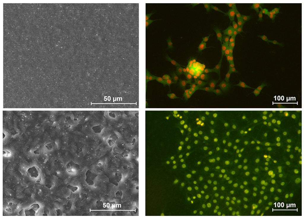
Share: