A new method for diagnosing cancer and metastases has been developed
16 December 2021 г. FRC KSC SB RAS

The spread of cancer cells through blood and lymph begins at the early stages of the disease. Sometimes this happens even before the primary tumor is large enough to be visualized with positron emission tomography, magnetic resonance imaging, or computed tomography. Traditional methods for determining the localization of metastases in the organism are neither sensitive nor specific enough to distinguish transformed small tissues. It is necessary to create a drug capable of selectively labeling tumor foci, even of critically small sizes, so that visualization could be performed on existing standard scanners.
A team of scientists from Siberia, including researchers from the Federal Research Center "KSC SB RAS", with the participation of colleagues from the University of Ottawa (Canada), developed a new radiopharmaceutical based on aptamers for the detection and visualization of cancerous tumors and metastases using positron emission tomography-computed tomography. This will significantly increase the accuracy of diagnosing and monitoring the development of cancer.
The new method uses anti-cancer DNA aptamers labeled with radioactive carbon to detect the localization of tumors and metastases. Aptamers are short oligonucleotide molecules synthesized in the laboratory which can bind to specific cells or other molecules. It is a cheaper and completely non-toxic alternative to antibodies for targeting, diagnosing, and delivering medicines. The carbon isotope used as a label is one of the fastest decaying and safe radionuclides which does not affect the chemical structure and properties of the biomolecule.
The resulting system was tested in mice with Ehrlich ascites carcinoma and its metastases. The radiolabeled aptamer made it possible to identify primary and secondary tumors smaller than two millimeters in size while the currently used radioactive glucose detects only seven millimeter foci. Specialists also note that such a combination of components provides highly specific and high-contrast images of cancer cells, does not give false positive results and can be used for targeted drug delivery.
“The identification of primary tumors and sites of metastasis is an important step in cancer diagnosis and monitoring of subsequent treatment. The synthesis of new radiopharmaceuticals for the treatment of cancer metastases is of great importance since the existing radionuclides for cancer imaging do not demonstrate sufficient selectivity, being accumulated in tumors and tissues with high metabolic activity. The advantages of aptamers as radioactively labeled probes for cancer imaging are target specificity and their rapid removal from the body without tissue damage. High specificity and affinity to the target receptor or cell, as well as the small size of the aptamers, provide good penetration into the tumor as well as good contrast image. For imaging, we labeled the aptamer with a radioactive carbon isotope with a short half-life. The resolution limit of standard radiopharmaceuticals is eight square millimeters. The carbon isotope aptamer has a much higher resolution, smaller than two square millimeters. We were able to distinguish small metastatic lesions throughout the organism: in thyroid gland, stomach, liver, kidneys, intestines, muscles, lungs, heart, pancreas, and even in bone marrow of the ribs. The created drug was completely removed from the body within an hour. The acute toxicity study showed that the aptamer-based pharmaceutical was not toxic. Consequently, this opens up new opportunities for cancer diagnostics, "- shared the results of the work Anna Kichkailo, Doctor of Biological Sciences, Head of the Laboratory of Digital Controlled Drugs and Theranostics of the Federal Research Center of KSC SB RAS, and Head of the Laboratory of Biomolecular and Medical Technologies of the V.F. Voino-Yasenetsky Krasnoyarsk State Medical University.
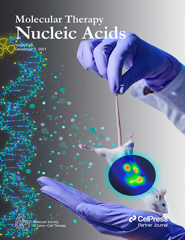
Note that the graphic representation of the research results of the Siberian scientists was used for the cover of the December issue of the journal Molecular Therapy - Nucleic Acids. Usually, the brightest and most interesting result is chosen as an illustration for the cover of a scientific journal.

Share: